Specialist Course on Light and Fluorescence Microscopy edition 2023
Description
Light microscopy, and especially fluorescence microscopy, is an essential tool to investigate and characterize biological specimen in the life sciences. Modern microscopes offer an increasing range of technical capabilities, but thereby become gradually more complex and require appropriate training for correct scientific use. This specialist course provides doctoral students with limited experience in microscopic imaging with the understanding that is needed to perform light and confocal imaging at a research-grade level.
Programme
- Day 1: Theoretical session (September 6 2023, 9-12 a.m.)
Lecture 1: Fundamentals of light and imaging – prof. Kevin Braeckmans
The main physical principles involved in image formation are explained. After reviewing concepts as light refraction, diffraction, resolution, etc., the basics of image formation are addressed. The differences between the types of objective lenses are discussed and attention is drawn to correct sample illumination.
Lecture 2: Transmitted light microscopy and digital imaging – Geert Meesen
The visualization of cells often proves to be difficult. The use of dedicated illumination techniques can enhance the image quality significantly. This lecture furthermore focuses on contrast-enhancing techniques such as DIC and phase contrast microscopy that help to reveal the smallest details in a cell. Concepts as resolution and noise are explored further and guidelines involving correct camera use and the impact of digital imaging are offered.
- Day 1: Demonstration sessions (September 6 2023, 1 – 5 p.m.)
Transmitted light microscopy – Dr. Herlinde De Keersmaecker, Geert Meesen
In the first demonstration session cell visualization with transmitted light microscopy is covered. The participants will learn in groups of 5 to set the correct sample illumination and explore different transmitted light microscopy techniques. The advantages of different contrast-enhancing techniques will be demonstrated and the principles of digital imaging are put into practice.
- Day 2: Theoretical session (September 7 2023, 9 – 12 a.m )
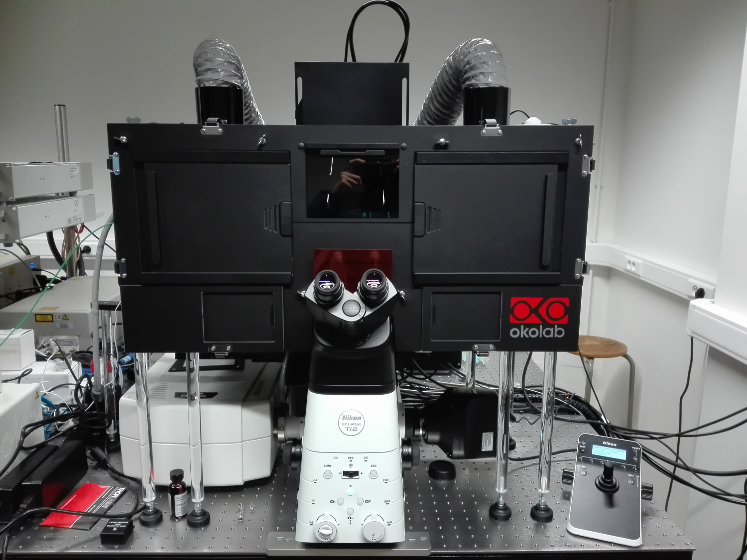
Lecture 1: Epi-fluorescence and confocal imaging – prof. Kevin Braeckmans
Fluorescence microscopy allows to image specific parts of a specimen with unrivalled contrast. In this session the basic concepts underlying fluorescence imaging are reviewed. It is explained how, with the use of various confocal techniques, optical slicing and three-dimensional imaging is achieved. Furthermore it is pointed out that the combination of confocal imaging and spectral detection is an excellent way to differentiate between different subunits in a sample. Special attention is drawn to practical considerations that are inherent to confocal microscopy, such as photobleaching and bleed-through.
Lecture 2: Advanced fluorescence microscopy: Strategies to image deeper or with higher resolution, a focus on two-photon imaging and STED nanoscopy: Dr. Deep Punj
In this lecture light is shed on two advanced light microscopy technique that are based on confocal laser scanning microscopy and are available at Ghent University. The first technique is two photon imaging, where the excitation strategy is shifted to higher wavelengths to allow sectioning as well as deeper penetration into tissue. The second technique is STED nanoscopy, with an adapted excitation scheme which allows to achieve resolutions up to around 40 nm, way beyond the optical resolution limit described by the Rayleigh criterion (~300 nm).
Lecture 3: Cell labelling and considerations for live cell imaging – Dr. Herlinde De Keersmaecker
This lecture explores the different properties of fluorescent labels and discusses the practical considerations when used for life science research. Different techniques used to introduce fluorescent dyes into a specific structure of both fixed and living cells are discussed. Attention is given to creating optimal conditions for live cell imaging.
- Day 2: Demonstration and peer assistant learning session (September 7 2023, 1 – 5 p.m.)
Epi- and confocal fluorescence imaging – Dr. Herlinde De Keersmaecker, Geert Meesen, Dr. Deep Punj, representative industry (Nikon)
The concepts of epi-fluorescence microscopy, confocal imaging and the two advanced techniques will be shown. Each session will demonstrate and focus on a specific capability and the related caveats of fluorescence imaging methods covered during the theoretical lectures with as goal to develop a correct scientific way of microscopic imaging. In between the demonstration sessions, two moments are foreseen to discuss and apply the new insights to a selection of their own research questions in small groups of 5 students. During the demonstrations time is foreseen to discuss questions from the peer assistant learning session related to the demonstration.
- Day 3: Demonstration and peer assistant learning session (September 8 2023, 9 – 11 a.m.)
Epi- and confocal fluorescence imaging – Dr. Herlinde De Keersmaecker, Geert Meesen, Dr. Deep Punj, representative industry (Nikon)
The concepts of epi-fluorescence microscopy and confocal imaging will be shown. Each session will demonstrate and focus on a specific capability and the related caveats of fluorescence imaging methods covered during the theoretical lectures with as goal to develop a correct scientific way of microscopic imaging. In between the demonstration sessions, two moments are foreseen to discuss and apply the new insights to a selection of their own research questions in small groups of 5 students. During the demonstrations time is foreseen to discuss questions from the peer assistant learning session related to the demonstration.
- Day 3: Peer assistant learning session (September 8 2023, 11 – 13 a.m.)
Each group of 5 students gives a 5-10 min presentation of the results of their discussion followed by a short Q&A session with all participants.
- Day 3: Guided computer session (September 8 2023, 14 – 17 p.m.)
Basics of image processing – Geert Meesen, Dr. Herlinde De Keersmaecker
Using a set of microscopy images, the fundamentals of image processing and quantitative image analysis are explained through a guided computer session. The session provides basic understanding and application of FIJI, an open source image processing platform widely used in biomedical research.
Remarks
Prices
Any discounted price depends of your profile (see shopping cart).
- UGent postDoc: 125 euro
- Non UGent non-profit: 250 euro
- Non UGent Profit: 900 euro
Teaching material
Access to a sharepoint platform for all digital documents (data sets for pc session, handouts of theoretical lectures, additional documentation) will be provided. Instrumentation of GLiM and microscopy demonstration samples will be available during demonstration sessions.
Organizing & Scientific Committee
Prof. Dr. Kevin Braeckmans
Faculty: Pharmaceutical Sciences - Department: Pharmaceutics
E-mail: Kevin.Braeckmans@UGent.be
Partners
Register here
Specialist Course on Light and Fluorescence Microscopy edition 2023 full programme
6 - 8 September 2023: Campus Heymans, Campus UZ, Campus Proeftuin
All participants will be notified prior to the start of the course with additional information regarding the course and exact locations.